Anti-PDCD1 Antibody, Rabbit Polyclonal
產(chǎn)品編號(hào):PA00169HuA10
$ 詢價(jià)
規(guī)格 50uL 100uL 200uL 可選
 |
說(shuō)明書(shū) |
產(chǎn)品名稱:Anti-PDCD1 Antibody, Rabbit Polyclonal
經(jīng)驗(yàn)證的應(yīng)用:WB
交叉反應(yīng):/
特異性:human PDCD1
免疫原:Recombinant human PDCD1 protein, fragment Pro21~Leu288; UniprotKB: Q15116
制備方法:Produced in rabbits immunized with human PDCD1, and purified by antigen affinity chromatography.
來(lái)源:Polyclonal Rabbit IgG
純化:Immunogen affinity purified
緩沖液:Supplied in PBS, 50% glycerol and less than 0.02% sodium azide, PH7.4
偶聯(lián)物:Unconjugated
狀態(tài):Liquid
運(yùn)輸方式:This antibody is shipped as liquid solution at ambient temperature. Upon receipt, store it immediately at the temperature recommended.
儲(chǔ)存條件:This antibody can be stored at 2℃-8℃ for one month without detectable loss of activity. Antibody products are stable for twelve months from date of receipt when stored at -20℃ to -80℃. Preservative-Free. Avoid repeated freeze-thaw cycles.
圖片:
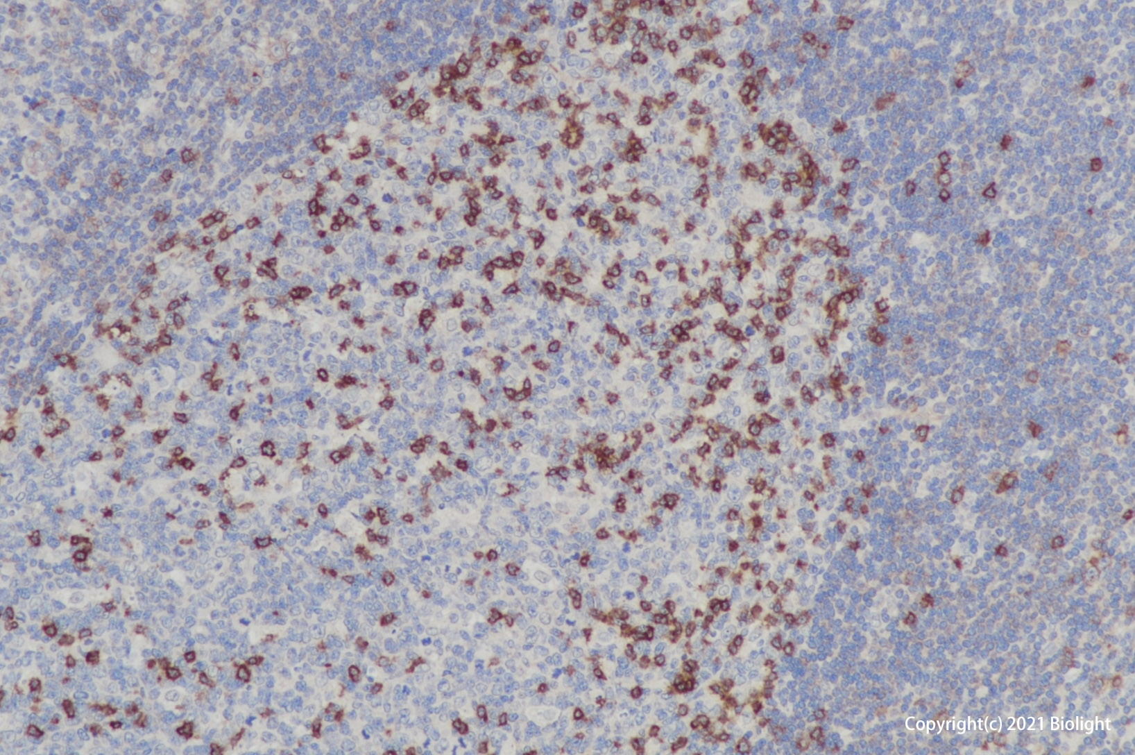
Figure1.Immunohistochemistry (Formalin/PFA-fixed paraffin-embedded sections) analysis of human tonsil sections labelling PDCD1 with purified PA00169HuA10 at 10ug/ml. Heat mediated antigen retrieval was performed using citrate buffer (pH 6.0). Tissue was counterstained with Hematoxylin. Rabbit specific IHC polymer detection kit HRP/DAB secondary antibody was used at 1/4000 dilution. PBS instead of the primary antibody was used as the negative control.
Figure2.Immunohistochemistry (Formalin/PFA-fixed paraffin-embedded sections) analysis of human lymphaden sections labelling PDCD1 with purified PA00169HuA10 at 10ug/ml. Heat mediated antigen retrieval was performed using citrate buffer (pH 6.0). Tissue was counterstained with Hematoxylin. Rabbit specific IHC polymer detection kit HRP/DAB secondary antibody was used at 1/4000 dilution. PBS instead of the primary antibody was used as the negative control.
別稱:CD279, PDCD1, SLEB2, HPD1P
背景信息:PD-1. Programmed Death-1 receptor (PD-1), also known as CD279, is type I transmembrane protein belonging to the CD28 family of immune regulatory receptors (1). Other members of this family include CD28, CTLA-4, ICOS, and BTLA (2-5). Mature human PD-1 consists of a 150 amino acid (aa) extracellular region (ECD) with one immunoglobulin-like V-type domain, a 21 aa transmembrane domain, and a 97 aa cytoplasmic region. The human PD-1 ECD shares 65% aa sequence identity with the mouse PD-1 ECD. The cytoplasmic tail contains two tyrosine residues that form the immunoreceptor tyrosine-based inhibitory motif (ITIM) and immunoreceptor tyrosine-based switch motif (ITSM) that are important for mediating PD-1 signaling. PD-1 acts as a monomeric receptor and interacts in a 1:1 stoichiometric ratio with its ligands PD-L1 (B7-H1) and PD-L2 (B7-DC) (6, 7). PD?1 is expressed on activated T cells, B cells, monocytes, and dendritic cells while PD-L1 expression is constitutive on the same cells and also on nonhematopoietic cells such as lung endothelial cells and hepatocytes (8, 9). Ligation of PD-L1 with PD-1 induces co-inhibitory signals on T cells promoting their apoptosis, anergy, and functional exhaustion (10). Thus, the PD-1:PD-L1 interaction is a key regulator of the threshold of immune response and peripheral immune tolerance (11). Finally, blockade of the PD-1: PD-L1 interaction by either antibodies or genetic manipulation accelerates tumor eradication and shows potential for improving cancer immunotherapy (12, 13).
全稱:Programmed cell death protein 1 (PDCD1)


